Your spine is the body’s central support structure which provides stability and flexibility while also protecting the spinal cord and nerves. The spine is comprised of several different components.
Vertebrae
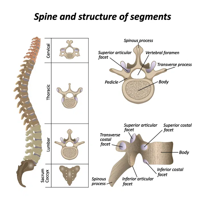
The spinal column is made up of 33 bones, called vertebrae, which are stacked on top of each other. The vertebrae provide structural support for the body but also protects the spinal cord, which runs through an opening in each vertebra called the vertebral foramen.
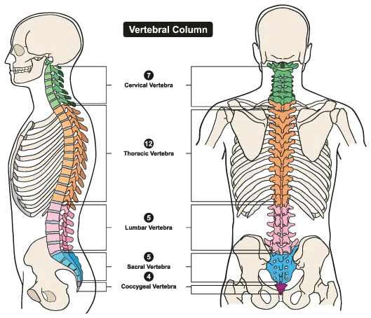
The vertebrae are divided into different regions based on their location in the spine:
- Cervical vertebrae: These are the vertebrae in the neck region and are numbered C1 to C7. They are smaller and more flexible than the other vertebrae, allowing for a greater range of motion in the neck.
- Thoracic vertebrae: These are the vertebrae in the mid-back region and are numbered T1 to T12. They are larger and more rigid than the cervical vertebrae and help to support the weight of the upper body.
- Lumbar vertebrae: These are the vertebrae in the lower back region and are numbered L1 to L5. They are the largest and strongest of the vertebrae and bear the majority of the body’s weight.
- Sacral vertebrae: These are the vertebrae in the pelvis region and are fused together to form the sacrum.
- Coccygeal vertebrae: These are the vertebrae at the very bottom of the spine and are also fused together to form the coccyx, also known as the tailbone.
Facet Joints
Facet joints are small joints located in the spine that help to provide stability and support while also allowing the spine to move and bend. They are found between the vertebrae, the bones that make up the spine, and are lined with cartilage to help them glide smoothly against each other. Facet joints are also known as zygapophyseal joints or Z-joints.
Discs
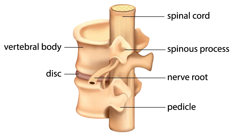
An intervertebral disc is a structure that sits between the vertebrae of the spine. It is made up of a tough outer layer of connective tissue called the annulus fibrosus and a softer, gel-like center called the nucleus pulposus. The intervertebral discs are important for the spine because they act as shock absorbers, allowing the spine to move and bend while also protecting the vertebrae from excessive force. They also help to distribute body weight evenly along the spine.
Spinal Cord and Nerves
The spinal cord is a long, thin, tubular bundle of nerves that extends from the brain down through the center of the spine. It is the main pathway for communication between the brain and the rest of the body. The spinal cord is encased in a protective sheath of connective tissue called the dura mater, which helps to protect it from injury.
The spinal cord is made up of millions of nerve fibers, which transmit signals to and from the brain. It is divided into 31 segments, each of which is connected to a pair of spinal nerves. These nerves branch out from the spinal cord and extend to the rest of the body, carrying sensory information to the brain and carrying instructions from the brain to the muscles and organs.
The spinal cord is an essential part of the nervous system and plays a vital role in the body’s ability to move, feel, and sense its surroundings. Damage to the spinal cord can have serious consequences, including paralysis and loss of sensation.
Soft Tissues
The soft tissues of the spine include a variety of structures that help to support and protect the spine. These tissues include:
- Muscles: The spine is surrounded by a number of muscles that help to support and stabilize the spine. These include the deep muscles of the back, such as the multifidus and the rotatores, as well as the larger muscles of the back, such as the latissimus dorsi and the erector spinae.
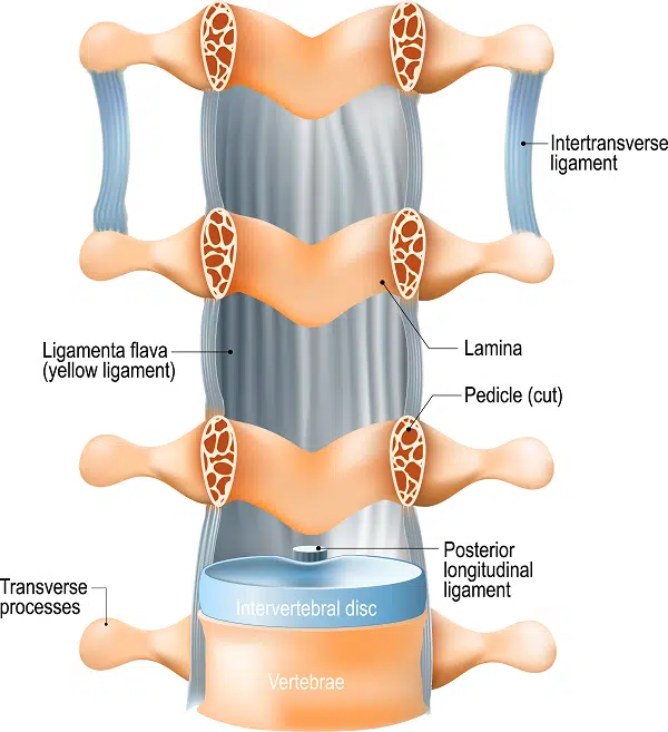
- Ligaments: Ligaments are strong bands of connective tissue that connect the bones of the spine to one another. They help to stabilize the spine and prevent excessive movement.
- Tendons: Tendons are strong, fibrous cords that attach muscles to bones. In the spine, they help to anchor the muscles to the vertebrae and allow them to move the spine.
- Fascia: Fascia is a type of connective tissue that surrounds and supports the muscles and other structures of the body. In the spine, it helps to protect the muscles and other soft tissues and provides a smooth surface for them to move against.
Together, these soft tissues help to support and protect the spine, allowing it to move and function properly.