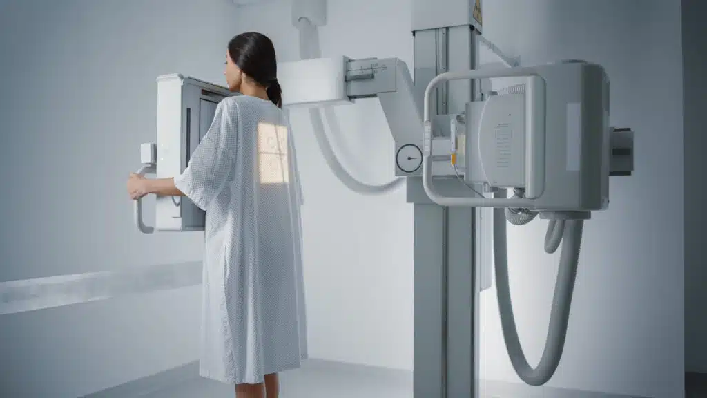
An X-ray image, also known as a radiograph, XR, or Xray, is a type of medical imaging that uses electromagnetic radiation in the form of X-rays to produce images of internal structures of the body. X-rays penetrate through the body, and different densities of tissue, such as bones and organs, absorb different amounts of the X-rays, creating an image on a film or digital detector.
How do X-rays work?
An X-ray machine produces a beam of X-rays, which are a form of ionizing radiation, by accelerating electrons through a high voltage. The electrons collide with a metal target, releasing X-rays in the process.
The X-rays penetrate through the body and pass through different tissues at different rates. Dense tissues such as bones absorb more X-rays and appear white on the resulting image, while less dense tissues such as muscles and organs absorb fewer X-rays and appear darker.
An X-ray film or digital detector is placed on the other side of the body from the X-ray source. The X-rays that pass through the body are recorded on the film or digital detector, creating a black and white image of the internal structures.
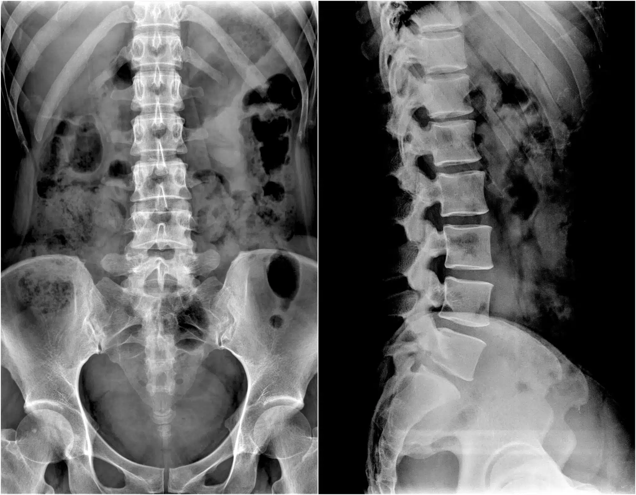
Which spine conditions can be diagnosed using X-ray?
X-rays are commonly used to diagnose problems with the spine. Some of the most common conditions that can be diagnosed with spinal X-rays include:
- Fractures or dislocations: X-rays can help diagnose fractures or dislocations of the vertebrae or other bones in the spine.
- Degenerative disorders: X-rays can help diagnose conditions such as osteoarthritis
- Scoliosis: X-rays can be used to diagnose scoliosis, a condition in which the spine has an abnormal curvature.
Benefits of X-ray
X-ray has a few benefits over other imaging studies which include:
- Cost: X-rays are often less expensive than MRI or CT scans.
- Availability: X-ray machines are widely available and typically faster to use, making X-rays a convenient option for many patients.
- Low radiation exposure: X-rays use low levels of ionizing radiation, which is generally considered safe for diagnostic purposes. CT scans, on the other hand, use higher levels of radiation, and repeated exposure can increase the risk of cancer.
- Real-time imaging: Some types of X-ray machines can produce images in real-time, allowing the doctor to see the movement of bones and joints, which can be useful in diagnosing certain conditions.
- Portability: X-rays can be performed at the bedside in a hospital or clinic, making them a convenient option for patients who are unable to move.
Limitations of X-ray
X-rays have some limitations compared to other imaging studies, such as MRI and CT scans. Here are some of the downsides of X-rays:
- Limited soft tissue detail: X-rays best produce images of bone and do not provide detailed images of soft tissues, such as muscles, nerves, and ligaments. For this reason, X-rays may not be the best option for diagnosing some conditions.
- Limited views: X-rays typically only produce images from one or two angles, making it difficult to fully evaluate complex conditions or structures.
- Radiation exposure: X-rays use ionizing radiation, which can increase the risk of cancer with repeated exposure. This may be a concern for some patients, especially women who are pregnant or trying to become pregnant.
In some cases, X-rays may be less effective than other imaging studies, such as MRI or CT scans, in diagnosing certain conditions. Your doctor will determine the best imaging test for you based on your symptoms and medical history.
Is X-ray safe?
X-rays are generally considered safe for diagnostic purposes, but like any medical procedure, there are some risks associated with exposure to ionizing radiation.
The risk of developing cancer from a single X-ray exposure is very low, but repeated exposure to ionizing radiation, as with multiple X-rays or other diagnostic tests that use radiation, can increase the risk of developing cancer.
Women who are pregnant should consider an alternative to X-ray if possible