Different imaging modalities, such as x-ray, CT, and MRI, are used to diagnose different medical conditions. Each imaging technology has its own set of strengths and weaknesses. For example, xrays, which are accessible and relatively inexpensive, do a good job detecting bone abnormalities but are inadequate to diagnose soft tissue problems. MRI does a better job of analyzing the soft tissues, but can be difficult to obtain given long wait times and high cost.
MRI, which stands for magnetic resonance imaging, uses a magnetic field and radio waves to create images of the body. The machine excites protons in the tissues of the body, and sensors measure their energy. Since protons are abundant in fat and water, MRI does a great job of visualizing tendons, ligaments, nerves, and intervertebral discs. This is why MRI is often considered the “gold standard” in diagnosing problems with the spine.
If you have undergone an MRI, understanding the results can be challenging. MRI is one of the more complicated imaging modalities to interpret, which is why a report is generally provided by a radiologist, who has undergone many years of medical and specialty training. Even these reports can be difficult to understand as they are typically written for other physicians and not usually intended to be read by patients directly. As a result, they typically contain esoteric medical terminology that’s unfamiliar to the average person.
Spine MRI Types and Views: T1s, T2s, Axial and Sagittal
An MRI image series is made up of various cross sectional “slices” or “cuts” that expose the anatomy various ways. You’ll notice that there are various sets of images with different contrast between tissues (sequences) and look at the body from different angles (views).
A sequence is a particular setting of the MRI which produces variance in the contrast for different tissues. Two of the most common sequences are T1-weighted and T2-weighted. Fatty tissues show up brighter in T1 whereas fatty and water-based tissues show up brighter on T2.
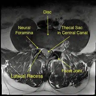
In addition to different sequences, there are various views of the body depending on the direction of the slices. The top down view of the body is known as the “axial” view, and a right-to-left view is known as the “sagittal” view.

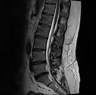
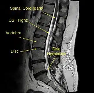
The sagittal view of the spine is useful for looking at the spinal column in totality. It allows you to evaluate the curvature of the spine as well as determine if there are any fractures or compression of the vertebral bodies. It is also helpful for examining the discs for degenerative changes and can be a helpful angle to see disc herniations protruding into the spinal canal.
The axial view provides a cross sectional view of the spinal canal and the tunnels through which nerve roots exit. You can better visualize certain parts of the vertebra and can identify disc herniations that may not be obvious in the sagittal view.