Magnetic Resonance Imaging (MRI) is a medical imaging technique used to visualize internal structures of the body in detail. It uses strong magnetic fields and radio waves to create detailed images of the body’s internal organs and tissues, including bones, muscles, and nerves.
How do MRIs work?
Magnetic Resonance Imaging (MRI) works by using strong magnetic fields and radio waves to generate images of the internal structures of the body. The basic principle behind MRI is that hydrogen atoms, which are abundant in the human body, can be aligned using a strong magnetic field and then disrupted by a radio wave pulse. The hydrogen atoms then emit a faint signal, which is picked up by detectors in the MRI machine and used to generate an image.
Which conditions are best diagnosed with MRI?
Magnetic Resonance Imaging (MRI) is a versatile imaging modality that can be used to diagnose a wide range of conditions. Here are some examples of conditions that are often diagnosed using MRI:
- Brain and spinal cord conditions: MRI is often used to diagnose brain and spinal cord conditions such as tumors, stroke, multiple sclerosis, and degenerative diseases such as Alzheimer’s and Parkinson’s.
- Joint problems: MRI can be used to diagnose problems with joints, such as osteoarthritis, ligament or cartilage tears, and inflammation of the joints.
- Abdominal and pelvic conditions: MRI is frequently used to visualize organs and tissues in the abdomen and pelvis, such as the liver, pancreas, kidneys, and uterus. It is often used to diagnose problems such as liver tumors, inflammation of the pancreas, and uterine fibroids.
- Breast conditions: MRI is sometimes used in addition to mammography to help diagnose breast cancer and other breast conditions.
- Cardiac and vascular conditions: MRI can be used to evaluate the heart and blood vessels, including the aorta, to diagnose conditions such as an enlarged heart, blockages in the blood vessels, and blood flow problems.
What to expect during an MRI?
Getting an MRI (Magnetic Resonance Imaging) scan is a non-invasive procedure that typically takes between 30 minutes to an hour. Here’s a general idea of what to expect when getting an MRI:
- Preparation: Before the MRI, you’ll be asked to remove any metal objects you’re wearing, such as jewelry, glasses, or hearing aids. You may also be asked to change into a hospital gown.
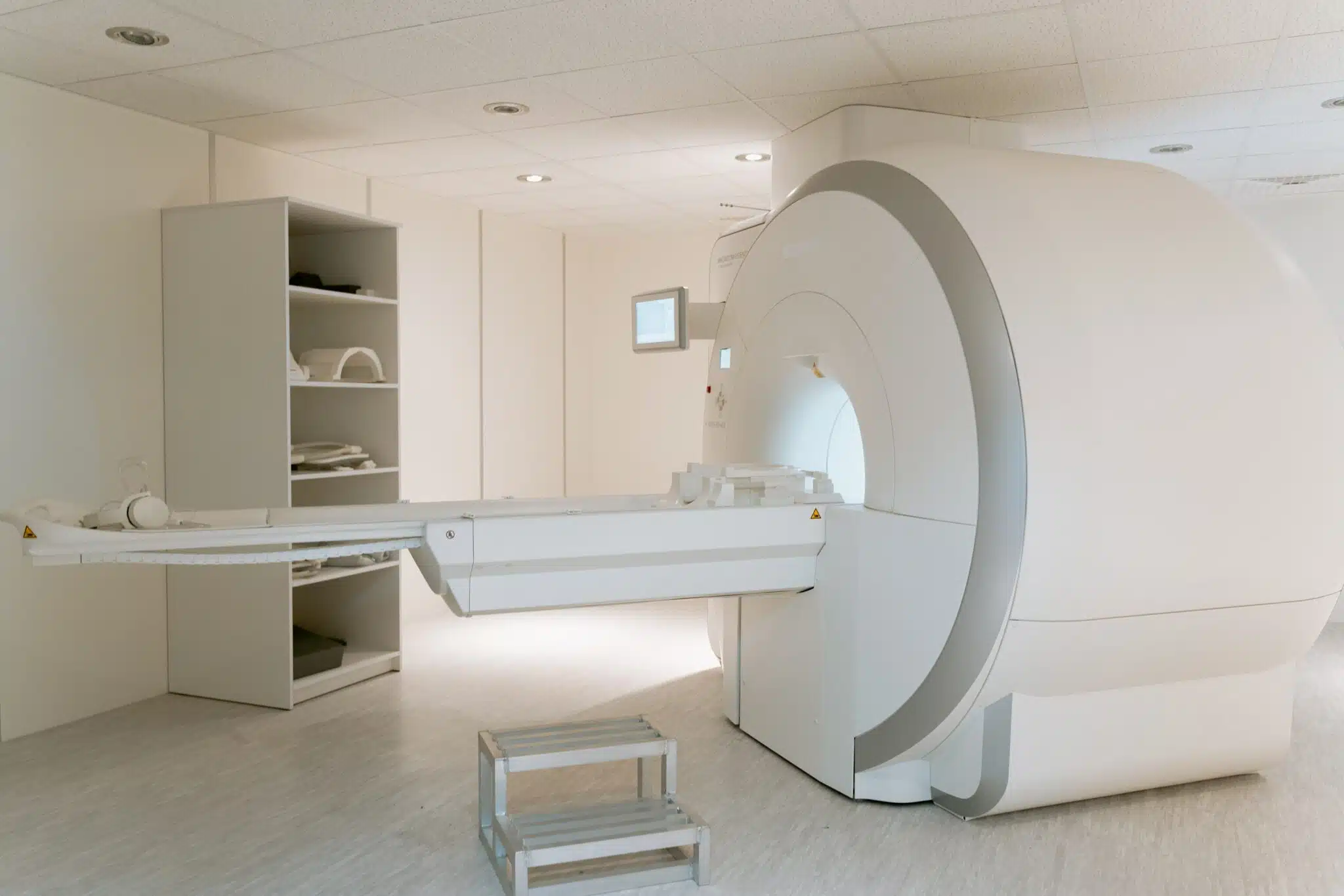
- Entering the machine: You’ll lie on a table that slides into a large cylindrical machine. Some people may feel a bit claustrophobic inside the machine, but you can ask for a larger bore or an open MRI if needed.
- The scan: Once inside the machine, you’ll be asked to lie still while the scan is performed. You’ll hear a series of loud thumping or knocking noises as the machine sends and receives signals. You’ll be given earplugs or headphones to help muffle the noise.
- The duration: The duration of the scan will depend on the type of scan you’re having and the area of the body being imaged. Some scans may take only a few minutes, while others can take up to an hour.
- After the scan: After the scan is complete, you’ll be able to get dressed and leave the facility. The images will be analyzed by a radiologist and the results will be sent to your doctor.
Are MRIs scary? Click here to learn more about what to expect and how to deal with issue such as claustrophobia.
It’s important to let your doctor know if you have any concerns or conditions that may affect your ability to lie still during the scan. For example, if you have a fear of enclosed spaces or suffer from anxiety, your doctor may be able to give you medication to help you relax during the scan. Additionally, if you’re pregnant or think you may be pregnant, it’s important to let your doctor know as some types of MRI scans may not be safe during pregnancy, such as those using contrast.
How long does an MRI take?
he duration of a Magnetic Resonance Imaging (MRI) scan can vary depending on several factors, including the area of the body being imaged. On average, an MRI scan can take anywhere from 30 minutes to an hour or more.
Here are a few different types of MRI scans and their typical duration:
- Head: 30-45 minutes
- Joint (e.g., knee, shoulder, hip): 30-45 minutes
- Spine (cervical, thoracic, or lumbar): 30-45 minutes
- Full-spine: 60 minutes or more
- Abdominal: 45 minutes to an hour
- Pelvic: 45 minutes to an hour
- Breast: 30 minutes to an hour
Note that these are just rough estimates and the actual duration of your MRI may be longer or shorter, depending on various factors.
Benefits of MRI
Magnetic Resonance Imaging (MRI) is a powerful imaging modality that has several benefits over other imaging methods, such as X-rays and CT scans. Some of the benefits of MRI include:
- No ionizing radiation: Unlike X-rays and CT scans, MRI does not use ionizing radiation, which can be harmful to the body. This makes MRI a safe imaging option for a wide range of patients, including pregnant women and children.
- Better visualization of soft tissues: MRI is particularly good at visualizing soft tissues, such as muscles, ligaments, and tendons, as well as the fat and fluid content in the body. This makes it an ideal imaging modality for diagnosing problems in these types of tissues.
- High-resolution images: MRI provides high-resolution images that can be very useful in detecting small changes or abnormalities in the body. This makes it an important tool for detecting conditions such as tumors, inflammation, and degenerative diseases.
- Versatile imaging modality: MRI can be used to visualize a wide range of tissues and organs in the body, including the brain, spinal cord, joints, abdomen, and pelvis. This versatility makes it a valuable tool for diagnosing a wide range of conditions.
Limitations of MRI
Magnetic Resonance Imaging (MRI) is a highly advanced imaging modality that provides detailed images of the body’s internal structures. However, like any imaging modality, it has certain limitations, including:
- Cost: MRI can be more expensive than other imaging modalities, such as X-rays and CT scans. This can be a barrier for some people who may not have insurance coverage or the financial resources to pay for the procedure.
- Time-consuming: MRI scans can take a significant amount of time, typically 30 minutes to an hour, which may be inconvenient for some people.
- Claustrophobia: Some people may experience anxiety or discomfort during the scan due to the enclosed space inside the MRI machine.
- Metal implants: People with metal implants, such as pacemakers, stents, or artificial joints, may not be able to undergo an MRI as the strong magnetic field can cause the metal to move or heat up.
Is MRI safe?
Magnetic Resonance Imaging (MRI) is generally considered safe for most people. Unlike X-rays and CT scans, MRI does not use ionizing radiation, which makes it a safer option for people who need to undergo repeated imaging studies or for those who are pregnant.
However, there are some safety considerations to keep in mind. People with certain medical devices, such as pacemakers, implantable cardioverter-defibrillators (ICDs), or cochlear implants, should not undergo MRI, as the strong magnetic field can interfere with the functioning of these devices.
People with metal implants, such as aneurysm clips or artificial joints, may also not be able to undergo MRI, as the magnetic field can cause the metal to heat up or move.
It’s important to discuss the benefits and limitations of MRI with your doctor and to carefully consider its use in light of your specific needs.