Neck pain and low back pain are common conditions affecting nearly 80% of people during their lifetime. Often the cause is from degenerative changes or minor trauma that will resolve on its own. However, there are many potential causes for neck or back pain, some more serious than others, so it is important to be evaluated by a medical professional.
History and Physical Examination
The first step in any evaluation is a thorough examination by a medical professional. This consists of an interview (known as history taking) as well as a physical examination.
Common questions that are asked during history taking include:
- Events surrounding the onset of pain (such as a fall)
- Quality of the pain (sharp, burning etc)
- Duration of the pain
- Associated symptoms (such as weakness or numbness)
- Factors that make it feel better or worse
A past medical history is also obtained to understand a patient’s risk factors for certain conditions.
The evaluation will also include a physical examination. Here the medical professional will examine the skin, press on the bothersome area, and have you twist and bend. They will also assess the strength and sensation in your arms and legs.
In most cases, the cause of spine pain can be diagnosed through history and physical examination alone.
Imaging Studies
If the pain is severe, lasts longer than expected, or has concerning features, additional studies may be warranted. Imaging studies are a common way of diagnosing the underlying cause of spine pain. Each visualizes different body parts and has its own strengths and weaknesses.
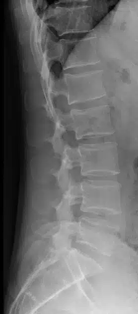
- X-ray – An X-ray uses the passage of electromagnetic waves to create shadow like images of bones and some organs. X-rays are quick and accessible but do a poor job evaluating soft tissues such as muscles and nerves. X-rays also expose the patient to a small amount of radiation.
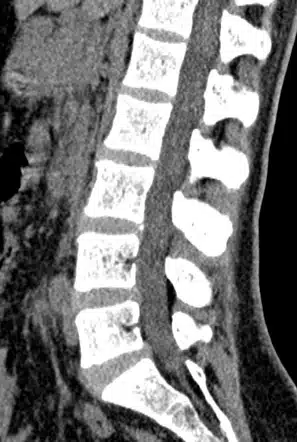
- CT scan – also known as Computed Tomography, this machine uses a series of x-rays along with computer processing to create detailed cross-sectional images. CT scans produce higher quality images that can also see organs and blood vessels. However, they are associated with more radiation exposure.
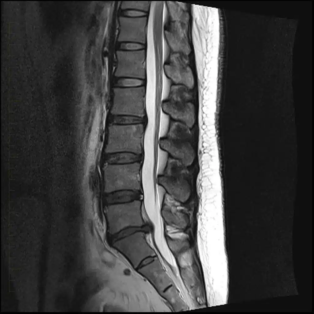
- MRI – which stands for Magnetic Resonance Imaging, uses a magnetic field and radio waves to create images of the body. MRI does a great job of visualizing tendons, ligaments, nerves, and intervertebral discs. Another benefit is that there is no radiation exposure. Unfortunately, MRI’s often are associated with longer wait times and higher cost. For more on MRIs, see How to Read a Spine MRI.
Lab Tests
Lab tests are sometimes ordered to help diagnose autoimmune disorders (such as rheumatoid arthritis or ankylosing spondylitis) or evaluate for signs of infection.
Electrodiagnostic studies (EMG/ECS)
Nerves and muscles produce electrical activity as they pass information to and from the brain. Electrodiagnostic studies can help determine if your symptoms are due to damage to a nerve or muscle. These tests are typically performed by a neurologist or a physiatrist (specialist in rehabilitation). The two most common electrodiagnostic tests are electromyography (EMG) and nerve conduction studies (NCS).
Electromyography (EMG)
An electromyography study (EMG) measures muscle response and electrical activity. During the test, a tiny needle is inserted through the skin into the muscle. The electrical activity is measured at rest as well as with muscle contraction.
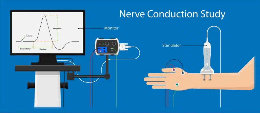
Nerve Conduction Study (NCS)
A nerve conduction study (NCS) measures the nerve and muscle’s response to an electrical impulse. In this study, electrodes are placed on the skin and mild electrical stimulation is administered to test the nerves ability to carry electrical impulses.
Diagnostic injections
A spinal injection can be used to help diagnose the cause as well as provide temporary relief. Numbing medication is injected into the site believed to be the cause of pain. A relief from pain indicates the potential origin.
Discogram
A discogram, also known as discography, is a test to determine whether the pain is originating from a specific intervertebral disc. This is an invasive procedure in which dye is injected into the disc under the guidance of a special real-time X-ray called fluoroscopy. If the pain is recreated, then this is a good indication that the particular disc is the source of pain.